|
|
|
 |
|
|
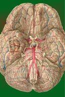
The white matter of the cerebrum is composed for the most part of the myelinated axons of neurons with cell bodies located in gray areas of the CNS. These gray areas include the cerebral cortex, thalamus, and basal nuclei (Figs-1 and 2). The basal nuclei along with the cerebellum play an important role in muscle coordination. They include the caudate nucleus, putamen, globus pallidus, and claustrum. However, the claustrum is often excluded when describing the functional role of the basal nuclei in motor control.
The putamen is continuous anteriorly with the head of the caudate nucleus, which arches upward and backward and finally curves around anteriorly and laterally to enter the temporal lobe ending in the amygdala (Fig-1). The caudate nucleus and putamen are collectively called the corpus striatum, while the putamen and globus pallidus represent the lenticular nucleus. Taken together, the caudate nucleus, putamen, and globus pallidus make up the basal nuclei.
The oval thalamic mass lies just medial and posterior to the lenticular nucleus on either side of the brain. With the exception of the auditory radiation from the medial geniculate body and the optic radiation from the lateral geniculate body, all ascending and descending pathways between the cerebral cortex and the brainstem pass through the internal capsule. This vertical group of fibers is bounded laterally by the lenticular nucleus, anteriomedially by the head of the caudate nucleus, and posteriomedially by the thalamus (Fig-2). The corona radiata is a fanlike radiation of ascending and descending fibers between the internal capsule and cerebral cortex. In cross section, the internal capsule forms a V -shaped region in each hemisphere (Fig-2). The principal ascending and descending fibers of the internal capsule are also illustrated in this figure. The anterior limb of the capsule is partly formed by the frontopontine fibers which arise in the cortex of the frontal lobe and descend to the pontine nuclei, where most of them synapse. The anterior limb also carries ascending fibers of the thalamocortical tract from the thalamus to the frontal cortex. Corticobulbar fibers from the motor cortex to cranial nerve nuclei controlling head and neck movements pass through the genu and anterior part of the posterior limb. Also located in the posterior limb, in increasingly posterior order, are corticospinal fibers to the limb and trunk muscles and the parietopontine and occipitopontine fibers connecting the parietal and occipital cortex with pontine nuclei. Most of the medial portion of the posterior limb is taken up by the thalamocortical fibers, which project to the parietal and occipital cortex.
|
|
|
Copyright [2007] [CNS Clinic-Jordan]. All rights reserved