|
|
|
 |
|
|
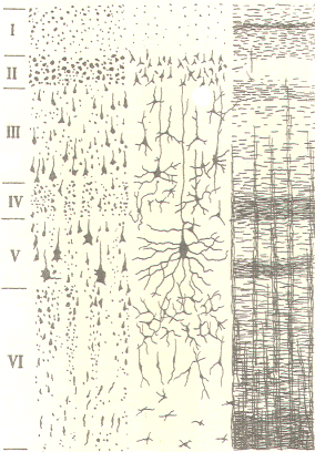
Phylogenetically, the human cerebral cortex is composed of a relatively recent and extensive portion, the neocortex, and an older relatively small region, the allocortex. The allocortex comprises only about 10 percent of the total cortical area and is limited to the olfactory cortex and the cingulate, parahippocampal and dentate gyri. It is functionally subordinate to the much larger neocortex, which comprises almost 90 percent of the cortex and represents almost all of the highly convoluted hemispheres seen in the exposed brain. The neocortex is composed of six distinguishable layers (laminae) which vary in thickness and density from one cortical region to another. The laminae are distinguishable from each other by the cell types found in each and by the type and direction of fibers which pass through them. The laminae are numbered from I to VI. with I being at the cortical surface and the others lying progressively deeper. The six laminae are described in Table -1.
Physiologists often subdivide the cerebral cortex into regions based on the functional characteristics of cortical layers in that region. Typically included are the sensory cortex (koniocortex), association cortex (homotypical cortex), and the motor cortex (heterotypical cortex). The sensory cortex includes the principal sensory receiving areas. while the association cortex covers major portions of the brain including the frontal, parietal, and temporal lobe. The motor cortex includes the principal motor areas. The relative thickness of cortical laminae IV and V is the noticeably variable feature between the three regions. The internal granular layer (IV) is the main receiving area for the sensory projection fibers from the thalamus. Consequently lamina IV is thickest in the sensory cortex. The internal pyramidal layer (V) is characterized by large pyramidal cells whose descending axons represent the motor fibers of the corticospinal system. Not surprisingly lamina V is largest in the motor regions of the cortex. Both laminae appear to be equally important in the association cortex as it receives some sensory input and gives rise to some motor output. It is also important to note that while the sensory cortex is primarily concerned with sensory input, it does give rise to a small motor component. Likewise the motor cortex receives a small degree of sensory input. The circuitry of the cerebral cortex has been much more difficult to evaluate than that of the cerebellar cortex. Because of the dense nature of neuronal elements, the extensive nature of dendritic processes called neutropil, and lack of repetitious patterns of neuronal contacts, meaningful evaluation of cortical neuronal circuitry has been difficult and not very fruitful. Recall that the neuronal makeup of the cerebellar cortex is everywhere identical and shows very symmetrical and repeated patterns. This, coupled with low neuronal density in the cerebellar cortex, has made experimentation and evaluation of cerebellar circuitry much easier than is true for the cerebral cortex. Another factor related to the difficulty of examining cerebral cortical circuitry is that fibers afferent to the cortex do not show the same consistency in their terminations as seen in the cerebellar cortex. Recall the climbing fiber-Purkinje cell and mossy fiber-granular cell synapses observed in the latter. Nevertheless, the efferent output from the cerebral cortex is primarily through axons of pyramidal cells in laminae II to V with the largest cells in lamina V. Cortical afferents project to all six laminae, with lamina IV of the sensory cortex receiving the largest number of collateral synapses.
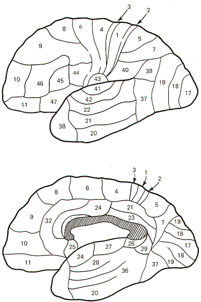
Brodmann, an early twentieth-century German neurologist, described the sixlayered cortex just discussed. Using Nissl stain, which clearly shows cell bodies but not neurites, he identified six distinct layers. Later work utilizing Golgi and Weigert stains brought out additional detail not previously seen. Brodmann mapped the cerebral cortex into many areas based on variations in the six layers. Many attempts have been made by physiologists to ascribe specific functional importance to these areas. In some cases this has been possible (e.g., Brodmann's area 17 and the primary visual cortex), but in many cases no distinct relationship exists. More often than not, specific functional regions seem to overlap several areas. Nevertheless, Brodmann's areas are quite useful as landmark indicators because of their worldwide recognition. The cortical areas of Brodmann are illustrated in the lateral and median sagittal views of Fig-1.
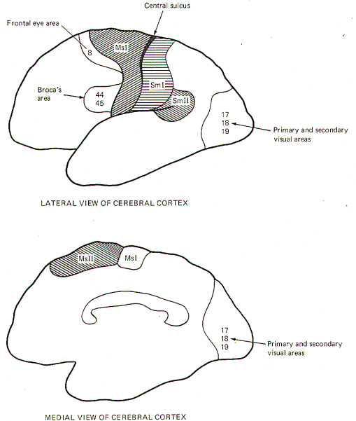
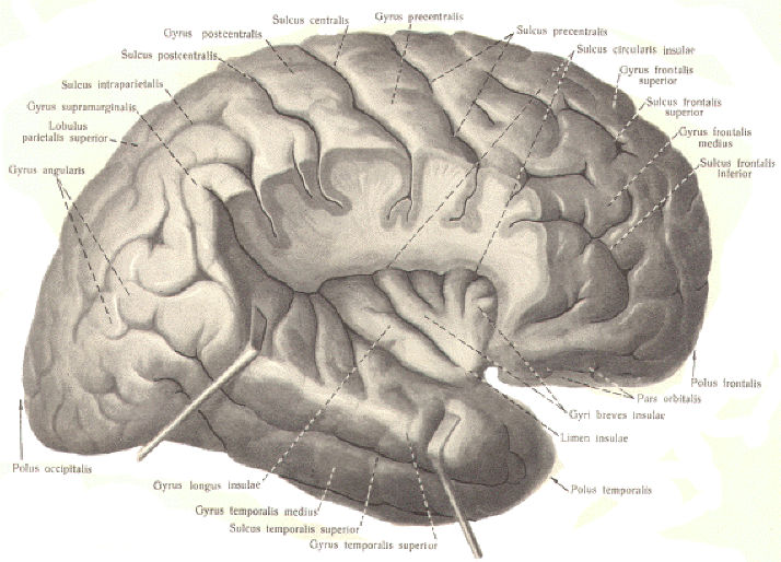
It is important to note that movements initiated in this way are not single uncoordinated contractions of given muscles, but rather movements accompanied by contraction of agonists and relaxation of antagonists. Nevertheless, these movements are very simple, and are similar to those which might be produced by an infant. Obviously more advanced movements must require the incorporation of additional systems. The primary motor area (equivalent to Brodmann's area 4 and an adjacent strip of area 6) extends over the superior medial border of the hemisphere onto the medial surface. The body is represented as a homunculus with the head and face regions located near the lateral fissure and the leg and foot areas extending onto the medial surface. The back extends anteriorly over area 4 onto the adjacent strip of area 6. The fingers and toes extend over the cortical surface in the central sulcus. Area MsI also has a small sensory component which receives input from a number of sources. The lemniscal system to the VPL nucleus of the thalamus ultimately projects from this nucleus to area 4 of MsI. The cerebellum projects to the VL nucleus of the thalamus, which in turn projects to areas 4 and 6 of MsI. Finally, the globus pallidus sends fibers to both the V A and VL nuclei of the thalamus which then project to area 6 of MsI. Much of the input to MsI is proprioceptive, but sensory input from other sources is also noted. The Supplementary Motor Area (MsII) The extension of area 6 onto the medial surface of the cortex represents the supplementary motor area (MsII). The body is represented horizontally here with the head forward, the back region lying adjacent to the cingulate gyrus, and the fingers just reaching the upper surface of the hemisphere. Electrical stimulation of this area produces somewhat complex bilateral avoidance movements. They are not as specifically distinct as those produced by MsI stimulation. The VA and VL nuclei of the thalamus both project sensory input to MsII. Both nuclei receive input from the globus pallidus, while the cerebellum projects only to the VL nucleus. The Frontal Eye Area: This region coincides with area 8. Electrical stimulation here produces deviation of the eyes, head, and neck to the opposite side. Broca's Motor Speech Area: This area corresponds roughly to areas 44 and 45 of the frontal lobe. Most of our information concerning its role comes from ablation studies and stimulation. Curiously, the left hemisphere appears to be dominant as ablation here usually abolishes sound production and often produces a motor aphasia or speech paralysis in humans. The effects typically aren't observed with ablation of the same area in the right hemisphere. It is estimated that 90 percent of us are left hemisphere dominant in this respect, regardless of right- or left-handedness.
The Primary Somatic Sensorimotor Area (SmI) Areas 3, 1, and 2 produce the largest evoked responses when somatic sensory receptors are stimulated. Smaller responses are recorded in the primary motor area (MsI) of the precentral gyrus. Input from touch, pressure, pain, temperature, and proprioceptors projects to the VPL nucleus of the thalamus, which then projects heavily to SmI, truly the principal receiving area for somatic sensation. The body homunculus represented in SmI is essentially a mirror image of that found in the primary motor area (Msl) anterior to the central sulcus. Studies with monkeys have indicated that the topographic organization of SmI consists of a series of overlapping bands at right angles to the central sulcus. Each of these bands is the cortical area for the representation of a single dermatome. It is also now well established that electrical stimulation of the postcentral gyrus (SmI) produces motor responses as well. It was originally thought that such movements were the result of current spread from the stimulating electrodes to the precentral gyrus. However, it is now quite clear that this is not the case and that SmI is capable of producing motor responses on its own, although requiring higher stimulation intensities than MsI. The Secondary Somatic Sensorimotor Area (SmII) This area, located immediately posterior to the face region of SmI, is characterized by a homunculus with the head represented anteriorly, the leg muscles posteriorly, the back inferiorly, and the hands and feet superiorly. Stimulation here causes gross movements of postural adjustments which are diffuse and widespread.
The Gustatory Cortex This area is located near the most inferior lateral extension of the primary sensorimotor area (Sml ) and may include area 43. Taste signals relayed through the VPM nucleus of the thalamus project to this area.
Certain behavioral patterns appear to be associated with one or the other hemisphere. These include handedness, the performance of speech, understanding the spoken and written word, and spatial appreciation. Approximately 90 percent of adults are right-handed (controlled by the left cerebral hemisphere) and over 96 percent of adults have their speech centers (Broca's motor speech area) located in the left hemisphere. This is evidenced by the fact that almost all aphasic patients with speech disorders have left-hemisphere lesions in Broca's area. Additional studies with humans have clearly established that the left hemisphere is best suited for written and oral language expression as well as analytic calculation, while the right hemisphere is particularly suited for appreciation of spatial relationships and aesthetic and nonverbal expression. Thus, the left hemisphere is often called the major or dominant hemisphere, while the right is the minor hemisphere. It should be noted that in those individuals who are left-handed, there is no similar shift in control from left to right hemisphere of the other behavioral observations listed above. Split-Brain Studies The commissural fibers (corpus callosum and anterior, posterior, and hippocampal commissures) serve to connect the two hemispheres. We have learned a great deal about the different functions of the two hemispheres from individuals who have had a complete sectioning of the corpus callosum in order to prevent the spread of epileptic seizures from one hemisphere to the other. These "split-brain" individuals retain normal behavioral patterns and can perform and learn as well as normal people. However, cerebral dominance shows up in carefully planned experiments. Much of this work has been performed by R. W. Sperry. If an unfamiliar object is placed in the left hand of blindfolded split-brain individuals, they will be able to fully appreciate its shape and touch by feeling it but will be unable to orally describe it or accurately draw a picture of it with their right hand. They cannot describe it orally because the right hemisphere, which received the sensory input from the object, is unable to communicate with the speech area of the dominant left hemisphere. Similarly, they will be unable to accurately draw it with the right hand because the important spatial information received by the right hemisphere cannot be transmitted to the dominant hemisphere. Thus, the apparent role of the cerebral commissures is the bilateral hemispheric integration of written and oral expression. Visual input to the split-brain individual likewise demonstrates cerebral dominance if carefully designed tests are performed. As the reader will recall, objects viewed in the left visual fields of both eyes are transmitted to the right occipital lobe. One such test involves having the individual look straight ahead at a table upon which are laid a variety of common objects such as a paper clip, screwdriver, bottle, key, etc. If a card bearing the printed name of one of these objects is flashed for 0.1 s in the left visual field, the individual is quite successful when asked to reach out and take the item named on the card with the left hand. This is so because the right hemisphere received the visual signal. and this same right hemisphere directs the movement of the left hand to the appropriate item. The subject could even crudely write the name of the retrieved item by writing with the left hand, since the right hemisphere, which received the signal, directs the activity of the left hand. However, because of the failure of the right (minor) hemisphere to communicate its information to the left (major) hemisphere because of the severed commissure, the individual cannot verbally say what name was seen on the card or what item was retrieved by the left hand. The left "speaking" hemisphere has not been informed of the actions of the right hemisphere. Indeed, the individual verbally denies even seeing such a card. Similarly, if asked to use the right hand to write the name of the item retrieved with the left hand, the subject would be unable to do so because the left "writing" hemisphere received none of relevant information. Identical results have now been observed in individuals who have their brains essentially "chemically split" by the injection of short-acting anesthetics into the left carotid artery, which anesthetizes the left hemisphere.
Much of our knowledge of the behavioral role of the various cortical areas has been obtained by accidental or disease-produced lesions in the human cerebral cortex. For example, lesions in areas 18 and 19 don't produce blindness, as visual signals still reach area 17 and objects can be clearly seen, but they are neither recognized identified, nor understood, This condition is known as visual agnosia, meaning that the viewed object is "not known." Lesions limited to area 17, however, produce outright blindness. Lesions to Broca's motor speech area (44 and 45) in the major hemisphere cause an expressive or motor aphasia. The patient can't speak intelligibly. He or she knows what to say but can't do it. There is no paralysis of the muscles themselves, but the patient speaks very slowly often leaving out nouns and verbs and has considerable difficulty with phrases. The caudal aspect of the superior temporal gyrus, known as Wernicke's area (area 22), is important for understanding the spoken word. Lesions to this area, typically in the major hemisphere, leave individuals able to hear normally, but spoken words appear to be meaningless. Such people can speak but make grammatical errors because of their failure to understand their own spoken words. This region receives many fibers from other association areas, including visual (18 and 19), auditory (41 and 42), and somesthetic (5 and 7). The condition, auditory aphasia, is most severe if the lesion involves both hemispheres, leaving these individuals unable to communicate orally in any intelligible fashion. The angular gyrus (area 39) is located at the caudal end of the lateral fissure between the supramarginal gyrus and Wernicke's area. Like Wernicke's area it has extensive connections with visual, auditory, and somesthetic association areas. A lesion of area 39 in the major hemisphere leaves the individual unable to comprehend written language. He or she can see words, but cannot understand them. This inability to read (alexia) does not prevent the individual from speaking normally but is usually accompanied by the inability to write (agraphia). Lesions to area 40, the supramarginal gyrus, inflicts a person with tactile and proprioceptive losses. This individual demonstrates astereognosis (the inability to identify familiar objects by touch) and makes errors in judgment concerning body position. While we can learn much about the contribution made by particular areas of the cerebral cortex from such lesion studies, it is important to recognize that the areas involved may simply be "links" in a chain as far as their affected sensorimotor observations are concerned. Other cortical areas may also be heavily involved. Further, eventually unraveling the sequences of cortical area involvement in a given behavioral pattern will still leave this greater mystery unsolved: What is the pattern of neuronal sequencing and synaptic integration?
Current research supports a theory that the motor cortex is composed of narrow, deep columns which represent the functional units around which sensorimotor activity is organized. These columns extend vertically through the entire six-layered cortex. Each may be as narrow as a single millimeter. Part of the basis for the columnar idea is based on observations that the cutaneous receptive field of a given column of cortical neurons lies in the path of movement produced by electrical stimulation of this same column. Thus as the muscles of a limb contract, objects encountered by the leading edge of the moving limb may possibly stimulate cutaneous receptors which reflexly reinforce the movement by projecting back to the same cortical column. Therefore, cutaneous inputs may be constantly providing feedback to guide limb movements via this corticomotor reflex. It is possible that this feedback may also involve muscle and joint receptors, causing the reflex to behave somewhat like a tracking system by directing the limb to follow the path of tactile and proprioceptive stimulation. The corticomotor reflex may also behave much like the stretch reflex in providing background muscle tone upon which the cortically originating voluntary motor command is superimposed. Each cortical column in the motor cortex is thought to have a facilitatory or inhibitory effect. Activity in horizontal association fibers from one cortical column to adjacent columns may provide the integration necessary for appropriate coordination of agonists and antagonists.
|
|
|
|||||||||||||||||||||||||||||||||||||||||||||||||||||||||||||||
Copyright [2007] [CNS Clinic-Jordan]. All rights reserved

