|  |  | 
				 Confusing 
				anatomy is often encountered during operations on complex 
				dysraphic lesions in the lumbosacral canal. It is common to see 
				nerve roots embedded in lipoma or scar tissue, or they may not 
				be easily distinguishable from fibrous adhesion bands. Sometimes 
				nerve roots that are bundled tightly by abnormally thickened 
				arachnoid can look like a thickened filum terminale. Also, the 
				transition between a functional but structurally deformed conus 
				and an intramedullary lipoma is not always visually apparent. 
				Thus, some objective means to identify the sacral nerve roots 
				and the conus is necessary to ensure preservation of these 
				neuronal structures. In addition, in some cases of complex 
				transitional lipomas, the tip of the conus is tautly suspended 
				by low sacral roots that are short, stout, and fibrotic. An 
				assessment of their functional integrity is useful for 
				determining whether dividing them, in order to complete the 
				untethering process, would lead to unacceptable loss of 
				sphincter function.  The first 
				sacral and lower lumbar roots are recognized readily by 
				intraoperative nerve stimulation while palpating for 
				contractions of the respective segmental muscle groups through 
				the surgical drapes. Identification of the lower sacral roots 
				and functional quantification of these roots and their 
				corresponding medullary connections, however, require some 
				objective assessment of perineal sensation and sphincter 
				function.  
				 Modality for Sensory Monitoring of S2-4 Segments The 
				assessment of evoked responses generated by directly stimulating 
				parts of the sex organs, urethra, and anal canal constitutes the 
				mainstay of sensory monitoring of the lower sacral segments. 
				Monitoring of such responses is most useful when the distal 
				conus or dorsal nerve roots are being rather strenuously 
				handled, as in certain difficult resections of large 
				transitional lipomas or during removal of the median fibrous 
				sleeve of a Type I split cord malformation. The latency and 
				amplitudes of the waveforms are exquisitely sensitive to 
				structural deformation and ischemic changes to the central 
				sensory pathways. Sensory evoked response monitoring is less 
				useful in the identification of sacral sensory roots, because 
				the responses are generated by end organ stimulation. Cortical 
				responses generated by direct dorsal root stimulation give much 
				less predictable waveforms, which are not stable enough for 
				foolproof identification purposes.  
				 Anatomy 
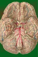
The 
				peripheral nerves that supply the bladder, anal canal, and 
				perineal skin, all potentially available for stimulation, are 
				divided into three main groups.  1.The 
				pudendal nerve is the primary somatic nerve to this region. The 
				pudendal motor neurons innervating the external sphincter and 
				pelvic floor originate from Onuf's nucleus in the anterior horn 
				of the S2 to S4 cord segments. The sensory fibers come from the 
				corresponding dorsal root ganglia. The mixed fibers course via 
				the S2, S3, and S4 roots to exit the spinal canal through the 
				sacral foramina (Figure-1). Somatosensory impulses travel in this nerve 
				from receptors located in the skin of the genitalia and 
				perineum, the pelvic floor, and bulbocavernosus muscles, as well 
				as in the mucosa of the distal urethra and anus. Motor fibers in 
				the pudendal nerve innervate the bulbocavernosus muscle, 
				external urethral sphincter, external anal sphincter, and pelvic 
				floor muscles. The pudendal nerve is the most easily accessible 
				nerve for evoked response testing.  Fig-1: 
				Schematic representation of the pudendal nerve and branching. 3.The 
				pelvic splanchnic nerves supply the sacral parasympathetic 
				innervation to the pelvic organs. The motor neurons in this 
				nerve originate in the S2 to S4 cord segments, slightly more 
				caudal than the pudendal motor neurons. The fibers are 
				distributed to the pelvic organs via the S2 to S4 nerve roots 
				and inferior epigastric plexus. The pelvic nerve carries sensory 
				afferents from the proximal urethra, bladder wall, prostate, 
				seminal vesicles, and rectum. Motor innervation is primarily to 
				the detrusor muscles, the corpus cavernosus, the rectum, and 
				probably the upper smooth-muscle portion of the external 
				urethral sphincter. Evoked responses can be elicited on 
				stimulation of the proximal urethra and bladder, presumably due 
				to activation of the pelvic sensory fibers.  3.The 
				hypogastric nerve plexuses carry autonomic (sympathetic) fibers 
				from the intermediolateral cell column of the T11-L2 spinal cord 
				segments. The preganglionic fibers course via the paravertebral 
				sympathetic chain ganglia, inferior mesenteric plexus, superior 
				hypogastric plexus, and finally the inferior hypogastric plexus. 
				The postganglionic fibers are distributed to the smooth muscles 
				of the bladder neck, the smooth-muscled internal urethral 
				sphincter, the parasympathetic intramural ganglia of the 
				detrusor muscles and probably the intrinsic portion of the 
				external urethral sphincter. The postganglionic fibers also 
				share connections with plexuses around the rectum and anal 
				canal, seminal vesicles, ductus deferens, prostate, and corpus 
				cavernosus in the male, and vagina in the female. It is 
				uncertain how much the afferent component of the hypogastric 
				nerves contributes to the evoked response in humans. 


							| 
				 Cortical Sensory Evoked Response 
							Standard recording of the cortical 
							evoked response is made by 5-mm silver or 
							gold-plated cup electrodes or dermal needle 
							electrodes sutured to the scalp. The electrode 
							impedance should be kept below 2000
							Ω. The active 
							recording electrode is placed in the midline, 
							approximately 2 cm behind the Cz 
							electroencephalographic recording site according to 
							the International 10-20 Electrode Placement System. 
							This has been demonstrated to give maximum cortical 
							response on stimulation of the penile and clitoral 
							skin. The reference electrode can be placed at a 
							number of sites, although the forehead (Fpz) is 
							convenient and gives a good waveform. Stimuli are 
							delivered at a rate of 3.5 to 5.0 per second, with 
							approximately 2.5 to 3.0 times the threshold 
							intensity. The recording console consists of high- 
							and low-frequency filters to keep the band pass at 
							30 to 1000 Hz. The sensitivity of the signal 
							amplifier is usually set at 2 to 10
							µV per division. 
							About 250 to 350 responses are averaged to ensure 
							reproducibility of the reading, but weak and 
							unstable signals from severely damaged conuses may 
							require up to 1000 responses to generate an 
							interpretable waveform. 
				 Pudendal Dermatomal Evoked Response 
							The most commonly used form of 
							pudendal nerve evoked response utilizes stimuli 
							applied to the sensory domain of the dorsal genital 
							nerve. In the male, the dorsal nerve of the penis 
							can be stimulated either bilaterally or unilaterally 
							using 5-mm cup electrodes placed 2 to 3 cm apart at 
							the base of the penis, with the cathode proximal to 
							the anode. Stimuli up to 3.0 or 3.5 times threshold 
							are well-tolerated. In the female, the dorsal nerve 
							of the clitoris is stimulated by 5-mm cup electrodes 
							or fine dermal needle electrodes fixed bilaterally 
							to the cleft between the labia major and labia 
							minor. The anodes are placed adjacent to the 
							clitoris bilaterally and the cathode approximately 2 
							cm posterior to the anode. 
							The averaged cortical pudendal evoked 
							response has a similar morphology as the responses 
							obtained from stimulation of the posterior tibial or 
							peroneal nerve. The response has a fairly 
							characteristic "M" pattern, with an initial positive 
							deflection followed by a constant negative, 
							positive, negative, positive waveform. Injury to the 
							S2-4 roots or cord segments is manifested by 
							lengthening of the P1 latency and 
							decreased amplitude of the triphasic waves (Figure 
							2). | 
							 |  
							| 
							Figure 2. Cortical pudendal nerve 
							evoked responses obtained from a male child with a 
							Type I split cord malformation. The neurological 
							deficits are much worse in the left leg. All 
							responses are recorded from Pz referenced to Fz.
							A)
							
							Tracing obtained by stimulating the right dorsal 
							nerve of the penis. B) Tracing obtained by 
							stimulating the left dorsal penile nerve. Note the 
							significant reduction in amplitude in the left 
							responses. |  


									|   
									
				 Urethral Evoked Response 
						Cortical evoked responses of very similar morphology and 
						latencies can be obtained using stimulating electrodes 
						embedded in a catheter inserted into the bladder. The 
						catheter has a balloon at its tip, which can be pulled 
						back snugly for anchorage. The location of the urethral 
						electrodes can be kept reasonably constant to eliminate 
						movement artifacts and interference.  
				 Anal 
						Evoked Response 
						Electrode-bearing catheters can also be inserted into 
						the anal canal for measurement of anal evoked responses. 
						The catheter is anchored by double balloons, the inner 
						one within the anorectal junction and the outer one 
						wedged at the anal verge. The cortical anal responses do 
						not differ from the urethral responses or the pudendal 
						dermatomal responses. 
				 Spinal 
						Evoked Response 
						Evoked responses can be recorded by electrodes placed on 
						the skin over the spine in humans. They reflect the 
						afferent volley traversing the dorsal columns. The 
						responses progressively increase in latency at more 
						rostral recording locations. Spinal evoked responses are 
						relatively easy to obtain in children, but the 
						amplitudes and waveform definition decrease with age, 
						such that by mid-teenage years, these responses are more 
						difficult to obtain, as in the case of adults. The 
						response over the mid-tolower lumbar spine consists of 
						an initially positive triphasic potential, representing 
						the volley as it ascends the cauda equina. Over the 
						caudal thoracic spine, the response consists of an 
						initially positive, predominantly negative triphasic 
						wave, the negative component of which has several peaks 
						or inflections. The initial portion of this response 
						arises in the intramedullary continuation of the dorsal 
						root fibers, and the subsequent portion reflects 
						synaptic activity concerned with local reflex mechanism 
						rather than the propagation of the response to more 
						rostral cord levels. From the mid-thoracic to the 
						cervical levels, the response consists of small, 
						triphasic potentials that are difficult to follow, 
						presumably arising from multiple ascending pathways 
						including the dorsal and dorsolateral columns. 
									
									The 
						only consistent spinal pudendal response has been from 
						stimulation of the dorsal nerve of the penis. The 
						recording electrodes are usually fixed at the T12-L1 
						interspinous space. The response has a morphology 
						comparable to the spinal response from the posterior 
						tibial and peroneal nerves but with smaller amplitudes 
						and a much shorter latency (Figure 3). The spinal 
						pudendal evoked response is sometimes not measurable in 
						overweight individuals, but its presence yields useful 
						information concerning peripheral sensory conduction 
						from the penis since it bypasses the central conduction 
						pathway rostral to the thoracic levels. Because the 
						cortical pudendal evoked response has similar latency 
						with the cortical posterior tibial response, the central 
						conduction time involved in the pudendal pathways must 
						be considerably longer than that in the posterior tibial 
						pathways. | 
									 |  
									| 
						Figure 3. Spinal pudendal evoked responses recorded over 
						the T12-L1 interspinous space on stimulation of the 
						dorsal nerve of the penis (upper), and spinal responses 
						on stimulation of the posterior tibial nerve (lower). 
						Note the much shorter latency of the pudendal response.
						 |  
				 Modality for Motor Monitoring of the S2-4 Nerve Roots 
						Pudendal sensory evoked responses are useful in 
						monitoring intraoperative injury to the conus and lower 
						sacral sensory nerve roots, but they are neither 
						qualitatively nor quantitatively suitable for the 
						identification of the lower sacral roots (especially the 
						motor roots) or conus from non-neural elements. 
						Intraoperative identification requires some way of 
						measuring the oneto-one stimulus-to-response coupling 
						of end organ function when the nerve root in question is 
						being stimulated. For the lower sacral roots, this means 
						the assessment of sphincter function.  
				 External Anal Sphincter Electromyography External anal sphincter electromyography (EMG) has long 
						been found useful as a qualitative tool for studying 
						anorectal closure function and disorders. The EMG 
						electrodes are either embedded in an anal plug or anal 
						balloon, which is placed into the anal canal, or are in 
						the form of needles inserted directly into the external 
						anal sphincter transmucosally. The needle electrodes are 
						more reliable because they are not subject to 
						dislodgement or to having mechanical artifacts during 
						contraction of the sphincter itself; however, accurate 
						and secure placement of the needles requires some 
						expertise and is initially best done by the 
						neuro-urologist. The grounding plate is pasted on the 
						patient's thigh, and EMG recordings are made using a 
						standard bladder diagnostic unit. The sensitivity of the recording stylus 
						is adjusted so that minimal deflection occurs at rest. 
						With stimulation of the lower sacral motor roots, the 
						stylus gives a discrete one-to-one spike-deflection, 
						much different than the baseline.  
						 
				 External Anal Sphincter Pressure Monitor The 
						external anal sphincter EMG requires bulky and expensive 
						equipment, as well as the availability of someone expert 
						in the accurate placement of the needle electrodes. An 
						alternate method of monitoring anal sphincter function 
						is the direct measurement of the "squeeze pressure" 
						induced by sacral root stimulation using a 
						pressure-sensitive balloon inserted in the anal canal. 
						This technique is simple and noninvasive, requires no 
						special expertise, utilizes inexpensive, portable 
						equipment, and produces easily interpretable pressure 
						waves which are semi-quantitative and virtually 
						unaffected by other electronic components in the 
						operating room that are known to cause annoying 
						baseline noise in an EMG recording. 
				
				 Physiology The 
						relationship between EMG and contractile strength in a 
						longitudinal muscle was first defined by Lippold, who 
						found a linear relationship between the integrated 
						action potentials on the EMG and the tension generated 
						by voluntary isometric contractions of the human 
						gastrocnemius. This linearity was explained by the fact 
						that an increase in contractile strength of 
						a muscle is brought about either by a spatially random 
						increase in the number of contracting motor units or by 
						random increments of discharge frequencies of the active 
						units; in both situations, the integrated electrical 
						output of the muscle would increase proportionately. The 
						same linear relationship was also demonstrated in the 
						external anal sphincter by Schweiger, who made 
						simultaneous recordings of sphincter EMG and anal canal 
						pressures with an anal balloon. These data support the 
						validity of using squeeze pressure, instead of sphincter 
						EMG, to monitor the functional status of the lower 
						sacral motor neurons. 
				 
						 In 
						order for the anal pressure monitor to be operational, 
						some sphincter function must be present. Theoretically, 
						a severely damaged motor nerve with only enough viable 
						axons to generate a barely visible EMG would produce no 
						measurable squeeze pressure; in such a situation, the 
						EMG might be more sensitive. However, such a nerve 
						would not provide useful sphincter function for the 
						patient, and its preservation is of doubtful value. In 
						the author's experience, any external anal sphincter 
						that could generate enough voluntary or reflex (as in 
						the bulbocavernosus or anal wink reflex in the infant) 
						contractions to be appreciable by preoperative digital 
						examination should produce recognizable pressure spikes 
						on the anal pressure monitor. This anal balloon monitor 
						is therefore sufficiently sensitive for the practical 
						purpose of sacral root and conus identification. 


				 Anatomy The 
						external anal sphincter consists of a bulky deep part, a 
						fusiform superficial part, and a subcutaneous part 
						decussating behind and in front of the anus. It 
						encloses the lower part of the levator ani, the 
						anorectal junction, and the anal canal in the shape of a 
						funnel. The internal anal sphincter arises from the 
						muscular coats of the rectum and insinuates itself 
						between the rectal mucosa and the upper portion of the 
						funnel. 
				 
						 The 
						external anal sphincter is innervated by the pudendal 
						nerve. This arises from the anterior division of S2 and 
						S3 and both divisions of S4, enters the pudendal (Alcock's) 
						canal through the lesser sciatic foramen, and divides 
						into two main branches just proximal to the urogenital
						diaphragm. The proximal branch, the inferior 
						hemorrhoidal nerve, supplies the striated muscles of 
						the external anal sphincter; the distal branch, the 
						perineal nerve, supplies the external urethral 
						sphincter. The internal anal sphincter, composed of 
						smooth muscles, is innervated by the hypogastric nerve, 
						derived from the intermediolateral (sympathetic) 
						columns of L1 and L2. Stimulation of the S2, S3, and 
						S4 roots, therefore, activates only the external and not 
						the internal anal sphincter. Furthermore, unless there 
						is localized disease or trauma to the pudendal branches 
						at the urogenital diaphragm, activity of the external 
						anal sphincter reflects function of the external 
						urethral sphincter. 
				 
						 The 
						anal pressure balloon described here is an elongated 
						ellipsoid selected specifically to pick up activities 
						from all three parts of the external sphincter funnel. 
						Its elongated span also minimizes the possibility of 
						accidental dislodgement by contractions of the pelvic 
						musculature induced intraoperatively. Although the 
						elongated balloon will also pick up contractions of the 
						internal anal sphincter, the latter is never activated 
						by the nerve stimulator or by manipulation of the lower 
						sacral spinal cord or nerve roots because its nerve 
						supply is from L1 and L2. However, being made up of 
						smooth muscles, the internal sphincter does have 
						spontaneous rhythmic contractions that will be 
						registered by the balloon, and these must be 
						distinguished from the stimuli-generated pressure 
						spikes from the external anal sphincter. 
							
								
									| 
				
				 Equipment The 
						pressure sensor is a double-lumen balloon catheter ordinarily used for 
						intraluminal angioplasty (Figure 4). The ellipsoidal 
						balloon is made of treated polyethylene, which does not 
						stretch or deform at high inflation pressures, so that a 
						high degree of sensitivity to circumferential squeezing 
						can be maintained. The central infusion catheter 
						concentric with the balloon is not actually being used 
						in the pressure measurement but functions effectively 
						as a stent for easy balloon insertion. The balloon comes 
						in different sizes, but the 3 x 0.8-cm (inflated 
						diameter) balloon should fit almost any patient, from 
						infants to large adults.    |  |  
									| Figure 4. Double-lumen polyethylene balloon catheter. 
						The infusion lumen is not involved in the pressure 
						measurement but merely serves as a stent. |  
							
								
									| 
									The 
						balloon is held vertically with the tip down, and is 
						maximally deflated and inflated several times with water 
						to expel all air bubbles. It is then connected to a 
						Bentley Model D240 pressure transducer, which displays the 
						pressure tracing on a two-channel Datascope Model 870 
									monitor (Figure 
									5). Although the baseline pressure of the balloon, which 
						can be adjusted by varying the amount of water used, 
						does not affect the actual pressure measurement, it 
						should be kept within a range that allows the monitor to 
						give good-sized pressure waves in the usual sensitivity 
						setting. The optimal condition is when the balloon is 
						rendered just turgid (with 0.8 ml water for the 3 x 
						0.8-cm balloon) and when the sensitivity on the Datascope 
									monitor is set at 25 (1 cm on the screen is 
						calibrated to 25 torr). The balloon is inserted into the 
						anal canal until its posterior end is just visible at 
						the mucocutaneous junction and then taped securely to 
						the gluteal skin. 
				 
						 One 
						cutaneous electrocardiography (ECG) electrode is pasted 
						over each iliac crest and a third on the right upper 
						thigh. The ECG tracing is displayed continuously on the 
						second channel of the Datascope screen (Figure 6). |  |  |  
									| Figure-5. Simple 
									assembly, consisting of the balloon 
									catheter, the Bentley pressure transducer, 
									the injecting syringe and stop-cocks and the 
									2-channel Datascope monitor. | Figure 6. ECG electrodes are placed over the iliac 
						crests and the thigh to register the stimulus artifact. |    
							
								
									| 
				
				 Technical Points 
						Intraoperative nerve stimulation is done with a 
						disposable monopolar nerve locator-stimulator using 3 V and three 
						variable current intensities: 0.5, 1, and 2 mA. The 
						monopolar stimulator is chosen over the bipolar 
						stimulator because only the former will generate 
						sufficient volume-conducted current to produce an 
						obvious stimulus artifact on the ECG when any tissue is 
						touched by the monopolar electrode. When a lower sacral 
						root is stimulated, the combined ECG stimulus artifact 
						and the pressure spike from the external anal sphincter 
						form an easily recognizable electromechanical couple on 
						the monitor (Figure 7).  
						There are two advantages in having this 
						electromechanical couple. 1) The stimulus artifact 
						eliminates the possibility of a faulty stimulator or 
						faulty stimulation technique because even at 0.5-mA 
						current, the monopolar electrode always produces a 
						prominent spike on the ECG tracing. If an artifact is 
						present without a corresponding pressure spike at high 
						current intensity, the tissue stimulated does not 
						innervate the external anal sphincter; if neither 
						stimulus artifact nor pressure wave is obtainable, then 
						the nerve stimulator is faulty (Figure 8). 
						 |  |  |  
									| Figure 7. Monopolar stimulations of the S3 root in a 
						5-year-old child. Note the prominent stimulus artifact 
						on the ECG tracing (arrows) coupled with large pressure 
						spike waves measured at 50-torr peak values. Scale in 
						torr. | Figure 8. Use of the EGG. A) Stimulation of a 
						nonfunctional element (fibrous adhesion band) showing 
						only EGG stimulus artifacts (arrows) but no pressure 
						response. Scale in torr. B) Recognition of a faulty 
						nerve stimulator when neither an EGG stimulus artifact 
						nor a pressure response is detectable. Scale in torr. |  
							
								
									| 
									2) 
						Involuntary, rhythmic activity of the internal anal 
						sphincter has been noted as spontaneous 5-to10-torr 
						waves with a frequency of 10 to 30 per minute, which 
						may be confused with external anal sphincter activities 
						except for the fact that these spontaneous waves are 
						completely out of phase with the ECG stimulus 
						artifacts (Figure 9). 
									During nerve stimulation, the cerebrospinal fluid must 
						be continuously suctioned away from the stimulation site 
						to prevent current dispersion. With this precaution, 
						supramaximal stimulation of the small sacral roots of 
						infants and young children can usually be accomplished 
						with 0.5 mA. The larger roots of adults sometimes 
						require higher amperage, as does direct conus 
						stimulation. Unilateral S2, S3, or S4 stimulation 
						consistently generates a peak pressure of 40 to 70 
						torr, in line with recordings reported by Lane of 60 to 
						125 torr pressures with voluntary (bilateral) 
						contraction in normal adults. Even in young infants, 
						peak pressure responses are generally above 40 torr. S3 
						stimulation produces the strongest and most consistent 
						response in the external anal sphincter. Direct 
						stimulation of the conus also results in waves of 
						comparable peak values but usually with a wider base, 
						probably because of multilevel and bilateral recruitment of anterior horn cell 
						units (Figure 10).  |  |  |  
									| Figure 9. Use of the EGG. Spontaneous lower pressure 
						waves «10 torr) from the internal anal sphincter are 
						completely out of phase with the EGG stimulus artifacts 
						(arrows). Scale in torr. | Figure 10. Direct stimulation of the conus in a 
						3-year-old boy, generating pressure waves with a wide 
						base and irregular blunted peaks. Stimulus artifacts are 
						indicated by the arrows. Scale in torr. |  
						Occasionally, a small pressure wave of less than 7 torr 
						follows S1 stimulation (which does not innervate the 
						sphincters) because of compression on the protruding 
						proximal portion of the balloon by the medial inferior 
						fibers of the gluteus maximus. Although such "ripple 
						waves" are easily differentiated from the tall spike 
						waves of healthy lower sacral roots, they could be 
						mistakenly construed as the subdued responses seen with 
						partially damaged S2, S3, and S4 roots. This confusion 
						is eliminated if care is taken to secure the posterior 
						end of the balloon just above the mucocutaneous 
						junction.  
						Stimulation of the filum terminale and nonneural 
						tissues always produces a stimulus artifact but not a 
						pressure wave. Thus, the S2, S3, and S4 roots and the 
						conus can be distinguished from the S1 and lumbar roots, 
						the filum, lipoma, fibrous adhesions, and other 
						nonfunctional fibroneural bands, such as an occult 
						myelomeningocele. The ECG artifact and pressure wave 
						relationships are summarized in Table 1. 
							
							
								| Table 1. 
								Interpretation of Stimulus (ECG) Artifact and 
								Pressure Response Relationship |  
								| Stimulus (ECG) Artifact | Pressure 
								Response | Interpretation |  
								| - | - | Faulty stimulator |  
								| + | -; spike waves 40-75 torr | S2,S3,S4, conus medullaris |  
								| + | - or ripple waves 
								(<7 torr) (and plantar flexion) | S1 |  
								| + | - | Lumbar roots, filum, non-neural 
								tissues |  
								| No stimulation | Rhythmic waves 10-30/min < 10 torr | Internal anal sphincter 
								spontaneous activity |    


								| 
					 Clinical Use of the Anal Sphincter Function Monitors The 
					anal sphincter function monitors (EMG or pressure balloon) 
					have been found useful in the following circumstances.
					 1. 
					Functional sacral nerve roots embedded in large lipomas may 
					be detected and traced through a sometimes aberrant course 
					to their exit foramina. This is particularly useful in 
					transitional lipomas that involve the dorsal as well as the 
					ventral portions of the conus 2. Sacral 
					nerve roots can be distinguished from fibrous adhesion 
					bands.  
					3. Atrophic, fibrous distal nerve roots in long-standing 
					myelodysplastic cases can be holding the conus tautly 
					against the dura and can thus prevent complete release of 
					the tethering. If these roots can be shown by the monitors 
					to have no contribution to sphincteric functions, they 
					should be cut.  
					4. Occasionally, the junction between functional conus and 
					fat is not well demarcated in cases of large transitional 
					lipomas or the type of terminal lipoma not having an 
					intervening filum terminale. Direct stimulation proceeding 
					from the obviously normal portion of the conus in a caudal 
					direction will identify the lowest extent of pudendal motor 
					neurons, beyond which sphincter contractions can no longer 
					be elicited by the nerve stimulator (Figure 11). | 
								 |  
								| Figure 11. Progressively caudal stimulation of the extremely 
				stretched-out conus of a 48-year-old patient with adult tethered 
				cord syndrome. Stimulation on the obviously normal portion of 
				the conus generated tall spike waves (A), stimulation at the 
				junctional zone between the conus and the filum produced smaller 
				waves with a wide base (B), and stimulation just beyond the 
				caudal extent of the conus elicited a minimal pressure response 
				(C). Arrows indicate stimulus artifacts. Scale in torr. |  
					 Pelvic Floor EMG Needle 
					recording electrodes can be percutaneously inserted into the 
					"extrinsic" portion of the external urethral sphincter to 
					monitor activity of this sphincter. This technique is 
					routinely used by neurourologists to correlate simultaneous 
					measurements of bladder pressure, urethral pressure, and 
					external urethral sphincter activities. Pelvic floor EMG can 
					thus be used for intraoperative sacral root identification 
					in the same manner as external anal sphincter EMG. 
					 
					 Modality for Sacral Reflex Monitoring Two 
					reflexes with centers in the sacral cord can be utilized to 
					assess the integrity of both the sensory and motor roots as 
					well as their interconnecting intramedullary components.
					 
					 Bulbocavernosus Reflex The 
					reflex response of the bulbocavernosus muscle to stimulation 
					of penile nerves can be studied using square wave electrical 
					stimuli applied through ring electrodes on the penis, and 
					recorded either by needle electrodes in the muscle or by 
					surface electrodes fixed to the midline of the perineum, 
					between the base of the penis and the anus. The averaged 
					response from 50 to 100 stimuli is usually biphasic with an 
					initial negative peak. The latency for most healthy adults 
					is 24 to 42 msec but varies with age and maturation in young 
					children. The waveform is also distorted significantly in 
					most cases of myelodysplasia and tends to become "unstable" 
					with very minor manipulations of the conus. The use of this 
					monitoring modality is therefore limited and is feasible 
					only in patients with virtually normal sphincter function 
					preoperatively.  
					 Urethral to Anal Sphincter Reflex Response The 
					urethral to anal sphincter reflex can be measured using 
					stimulating electrodes similar to those used in eliciting 
					urethral cortical evoked responses and recording electrodes 
					used in recording external anal sphincter EMG. The latency 
					is considerably longer (50 to 70 msec) than the 
					bulbocavernosus reflex, although their morphologies are 
					similar. The long latency in the urethral-anal sphincter 
					reflex is due partly to the slower conducting velocity of 
					autonomic afferent fibers and partly to a more complex 
					central polysynaptic reflex organization.   |  | 
					
						|  |  
						| 
							
								| 
	
		| Our brain is a mystery and to understand it, you 
		need to be a neurosurgeon, neuroanatomist and neurophysiologist. 
		 neurosurgery.tv
  Please visit this site, where daily neurosurgical activities are going 
		on.
 
		 Inomed ISIS IOM System
 |  |  |  |